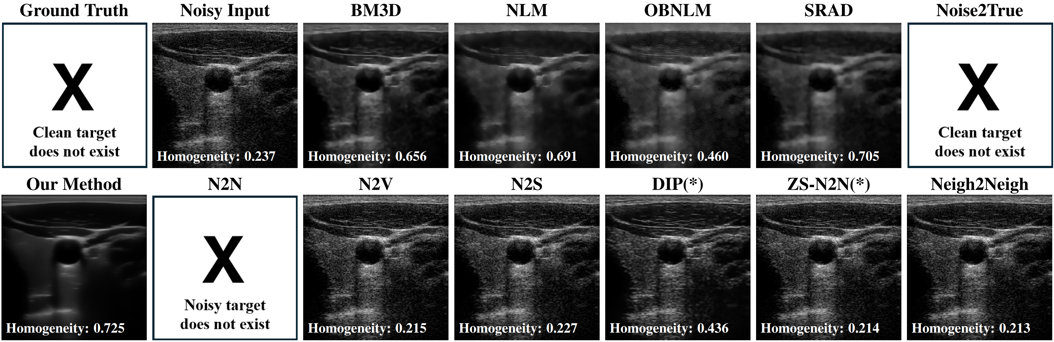
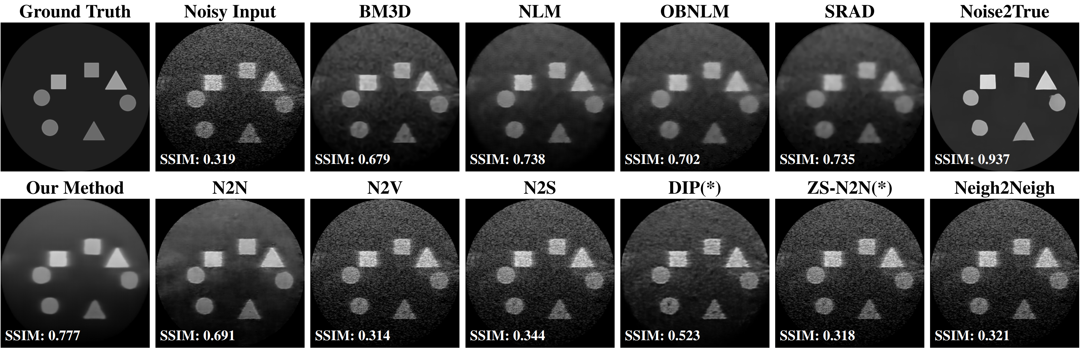
Swipe the figures sideways to see every method.
Medical Image Analysis · 2025
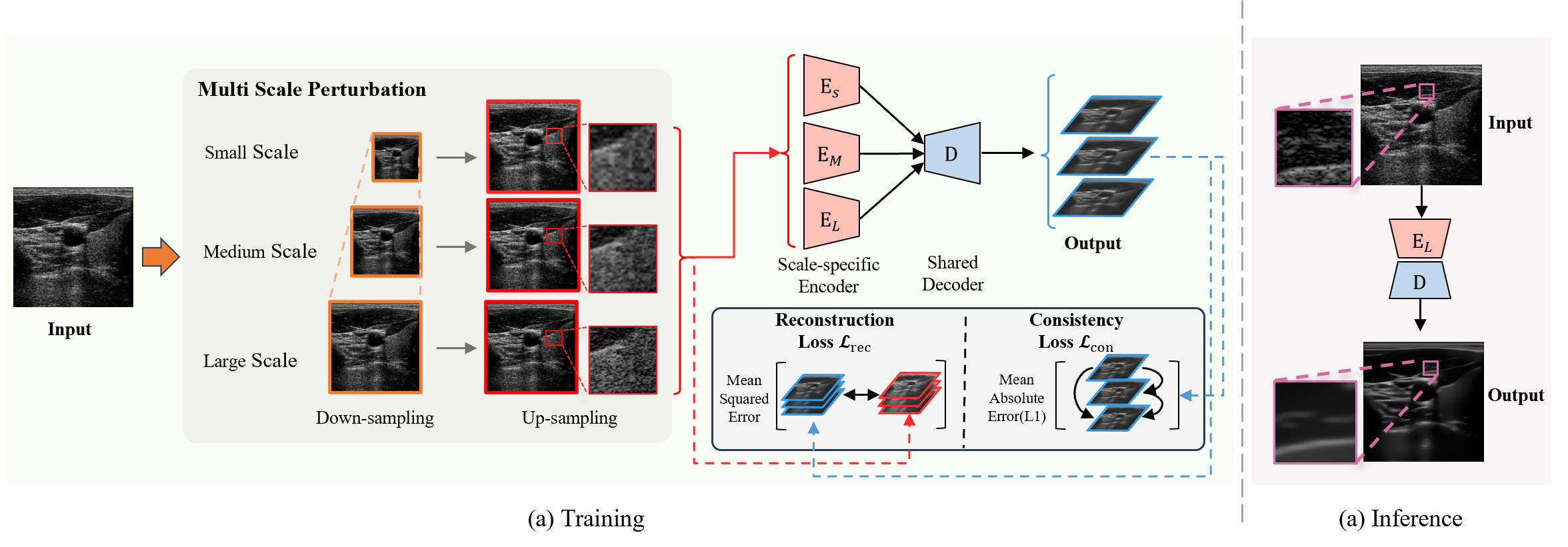
Image denoising is a fundamental task in computer vision, particularly in medical ultrasound (US) imaging, where speckle noise significantly degrades image quality. Although recent advancements in deep neural networks have led to substantial improvements in denoising for natural images, these methods cannot be directly applied to US speckle noise, as it is not purely random. Instead, US speckle arises from complex wave interference within the body microstructure, making it tissue-dependent. This dependency means that obtaining two independent noisy observations of the same scene, as required by pioneering Noise2Noise, is not feasible. Additionally, blind-spot networks also cannot handle US speckle noise due to its high spatial dependency. To address this challenge, we introduce Speckle2Self, a novel self-supervised algorithm for speckle reduction using only single noisy observations. The key insight is that applying a multi-scale perturbation (MSP) operation introduces tissue-dependent variations in the speckle pattern across different scales, while preserving the shared anatomical structure. This enables effective speckle suppression by modeling the clean image as a low-rank signal and isolating the sparse noise component. To demonstrate its effectiveness, Speckle2Self is comprehensively compared with conventional filter-based denoising algorithms and SOTA learning-based methods, using both realistic simulated US images and human carotid US images. Additionally, data from multiple US machines are employed to evaluate model generalization and adaptability to images from unseen domains.
Drag the slider to compare the results of different despeckling methods side by side. Use the frame slider to select different B-mode slices.
If the views become unsynchronized after switching methods, please switch to another frame to refresh.
Quantitative comparison of different methods for US speckle reduction in terms of PSNR, SSIM, Homogeneity (Hom), and LPIPS. The best-performing method for each metric, trained without clean images, is highlighted in bold. The supervised method results are marked in a gray shade. Note that for clinical carotid images, the Noise2Noise and supervised Noise2True methods are not applicable.
| Method | S-I Data | S-II Data | S-III Data | Carotid | Time | |||||||||
|---|---|---|---|---|---|---|---|---|---|---|---|---|---|---|
| PSNR ↑︎ | SSIM ↑︎ | Hom ↑︎ | LPIPS ↓︎ | PSNR ↑︎ | SSIM ↑︎ | Hom ↑︎ | LPIPS ↓︎ | PSNR ↑︎ | SSIM ↑︎ | Hom ↑︎ | LPIPS ↓︎ | Hom ↑︎ | ||
| Non-learning based | ||||||||||||||
| SRAD | 19.40 | 0.734 | 0.763 | 0.411 | 19.57 | 0.735 | 0.768 | 0.419 | 20.81 | 0.880 | 0.874 | 0.246 | 0.705 | 6 s |
| NLM | 19.40 | 0.736 | 0.714 | 0.417 | 19.52 | 0.738 | 0.740 | 0.428 | 20.94 | 0.881 | 0.839 | 0.310 | 0.691 | 80 ms |
| BM3D | 19.26 | 0.684 | 0.691 | 0.397 | 19.34 | 0.679 | 0.694 | 0.407 | 21.01 | 0.875 | 0.844 | 0.245 | 0.656 | 3 s |
| OBNLM | 18.84 | 0.703 | 0.577 | 0.470 | 18.88 | 0.702 | 0.621 | 0.484 | 21.49 | 0.885 | 0.774 | 0.299 | 0.460 | 12 s |
| Supervised (Noisy-Clean Pairs) | ||||||||||||||
| Noise2True | 23.33 | 0.936 | 0.910 | 0.059 | 23.25 | 0.937 | 0.923 | 0.049 | 27.66 | 0.973 | 0.941 | 0.062 | × | 6 ms |
| Self-supervised (Noisy-Noisy Pairs) | ||||||||||||||
| N2N | 18.05 | 0.705 | 0.683 | 0.369 | 17.74 | 0.691 | 0.689 | 0.410 | 21.99 | 0.895 | 0.820 | 0.240 | × | 6 ms |
| Self-supervised (Single Noisy Images) | ||||||||||||||
| DIP(*) | 18.87 | 0.543 | 0.515 | 0.484 | 18.93 | 0.523 | 0.531 | 0.501 | 20.98 | 0.831 | 0.747 | 0.323 | 0.436 | × |
| N2V | 17.62 | 0.311 | 0.312 | 0.561 | 17.55 | 0.314 | 0.339 | 0.551 | 19.87 | 0.613 | 0.573 | 0.388 | 0.215 | 6 ms |
| N2S(*) | 17.32 | 0.303 | 0.308 | 0.549 | 17.64 | 0.310 | 0.339 | 0.545 | 19.69 | 0.587 | 0.576 | 0.399 | 0.227 | × |
| N2S | 18.14 | 0.346 | 0.316 | 0.543 | 17.64 | 0.344 | 0.330 | 0.547 | 20.41 | 0.657 | 0.573 | 0.399 | 0.184 | 6 ms |
| Neighbor2Neighbor | 17.63 | 0.319 | 0.310 | 0.556 | 17.79 | 0.321 | 0.339 | 0.549 | 20.19 | 0.637 | 0.574 | 0.384 | 0.213 | 6 ms |
| ZS-N2N(*) | 17.58 | 0.315 | 0.312 | 0.557 | 17.71 | 0.318 | 0.340 | 0.550 | 20.04 | 0.625 | 0.576 | 0.384 | 0.214 | × |
| Speckle2Self (our) | 18.59 | 0.771 | 0.803 | 0.231 | 18.94 | 0.777 | 0.792 | 0.233 | 22.90 | 0.922 | 0.876 | 0.173 | 0.725 | 7 ms |
Swipe the table sideways to see every column. View the original table image.
@article{li2025speckle2self,
  title={Speckle2Self: Self-supervised ultrasound speckle reduction without clean data},
  author={Li, Xuesong and Navab, Nassir and Jiang, Zhongliang},
  journal={Medical Image Analysis},
  pages={103755},
  year={2025},
  publisher={Elsevier}
}